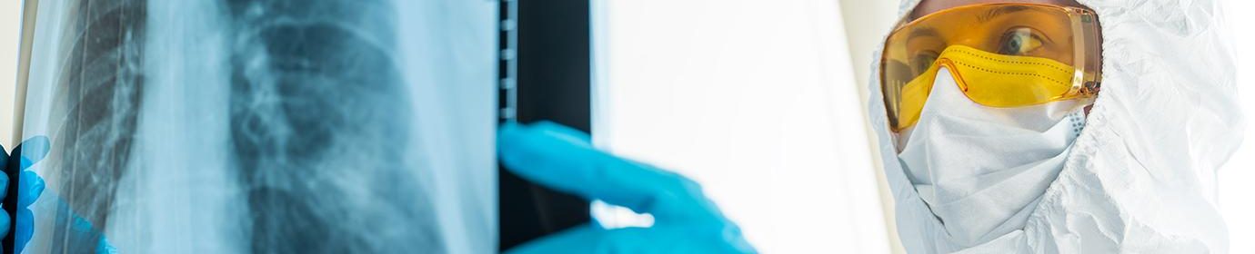
Researchers Develop AI to Diagnose, Understand COVID-19 in Lung Images
UPDATE: Maryellen Giger was also chosen to lead a new NIH-funded Medical Imaging and Data Resource Center (MIDRC), which will create an open source database with medical images from thousands of COVID-19 patients.
As physicians and researchers grapple with a rapidly-spreading, deadly and novel disease, they need all the help they can get. Many centers are exploring whether artificial intelligence can help fight COVID-19, extracting knowledge from complex and rapidly growing data on how to best diagnose and treat patients.
One University of Chicago and Argonne National Laboratory collaboration believes that AI can be a helpful clinical partner for a particularly important kind of medical data: images. Because severe cases of COVID-19 most often present as a respiratory illness, triggering severe pneumonia in patients, chest X-rays and thoracic CT scans are a potential exam. With a grant from the new c3.ai Digital Transformation Institute, computer-aided diagnosis expert Maryellen Giger will lead an effort to develop new AI tools that use these medical images to diagnose, monitor and help plan treatment for COVID-19 patients.
Once developed, Giger hopes that the system will help clinicians in several different aspects of diagnosing and treating the disease. First, it potentially could aid radiologists in spotting the disease, both in patients already suspected of COVID-19 and in those who undergo lung screening for other conditions. Second, the AI model could help physicians differentiate between different stages of the infection, guiding treatment choices for individual patients. As time goes on, researchers also hope the new AI method could help identify cases of COVID-19 that were missed due to lack of symptoms or unavailability of tests.
“There are multiple parts: the detection and diagnosis, as well as the response to treatment,” Giger said. “How do we follow the disease? And when do we see a presentation that says ‘start treatment’? And if they are being treated, how can we follow to determine if the treatment is working? Can we use this to inform decisions and treatment?”
The project builds upon Giger’s decades of experience in developing AI methods to analyze medical images and interpret signs of disease, including lung, breast, and prostate cancer, lupus, and thyroid cancer. The research will draw upon approaches in computer vision and deep learning from her UChicago lab, clinical lung imaging expertise from the UChicago Department of Radiology, and computational resources at Argonne.
“We’re taking our AI development knowledge that we’ve learned from analyzing other lung diseases and using it here,” Giger said. “In doing so, we will conduct transfer learning and get away with a lot fewer cases as we combine medical imaging intelligence with machine intelligence.”
Like other clinical computer vision systems, the COVID-19 tool will be trained with real data to differentiate between chest images from patients with and without the infection. But this training typically requires large amounts of data, and in these early stages of the pandemic, COVID-19 medical images following patients from diagnosis through recovery remain scarce. The project will begin with a dataset from Chinese patients, and expand to include cases from UChicago Medicine and other health centers as it proceeds to a goal of 1000 cases.
But it will also take advantage of their cutting-edge computational method known as cascade-based deep transfer learning to speed up this process. Instead of starting from scratch, they will start with network models Giger’s lab has developed for assessing other lung conditions and gradually retrain them to detect COVID-19 hallmarks. The approach carries clinical logic as well, as different stages of COVID-19 may present similar features to other lung diseases, such as cancer and other pneumonias.
Some of the image features used by the model in assessing severity of disease could also serve as important image-based biomarkers for use by clinicians to monitor disease progression or recovery.
Eventually, researchers hope the new AI method could help identify cases of COVID-19 that were missed due to lack of symptoms or unavailability of tests. Parallel AI methods from Giger’s group have looked at the “incidental” detection of emphysema, heart disease, and osteoporosis from thoracic CT scans gathered during lung cancer screening, as well as mesothelioma within her colleague’s lab. As coronavirus spreads through the population, these medical images may also reveal telltale signs of past or present COVID-19 infection, enabling treatment and providing valuable public health information.
“The impact of this research area is expected to be great because, as we move into a potential next wave or if COVID is looked at as a chronic disease, we will need to be able to recognize it,” Giger said. “One of the ways of doing that is through what’s routinely obtained: chest radiographs and low dose CTs for lung screening. So this will just be another checkbox of ‘is there COVID potential here?’”
The project was awarded a grant as part of the first call for proposals from the new C3.ai Digital Transformation Institute, a partnership between the University of Chicago and five other research universities with tech companies C3.ai and Microsoft. Additional grant recipients will develop new mathematical models for predicting the spread of COVID-19 and the effectiveness of mitigation policies, and study the effect of the pandemic on housing security and inequality.
“These first three research projects represent the breadth of solutions for COVID-19 mitigation that artificial intelligence can bring to bear on fields as disparate as medicine, urban planning, and public policy,” said Thomas M. Siebel, CEO of C3.ai. “Through the C3.ai Digital Transformation Institute, we have the opportunity to change the course of a global pandemic.”
The UChicago project also includes Jonathan Chung, Vice Chair of Radiology for Quality and Section Chief of Cardiopulmonary Imaging at UChicago Medicine; Samuel Armato, Associate Professor of Radiology and Chair of the Committee on Medical Physics at UChicago Medicine; Ravi Madduri, Computational Scientist in the Mathematics and Computer Science division at Argonne; and Hui Li, Research Associate Professor in the Department of Radiology at UChicago Medicine.
DSI Software Engineers create interactive map tool to maximize climate investment tax benefits

Transform cohort 3 participant Healee uses AI to improve healthcare
Haifeng Xu named a AI2050 Early Career Fellow